
A simple test that monitors the electrical impulses generated around your heart.
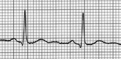
Electrodes are attached to the torso that precisely measure the bio-electric voltage in your heart. The result is a simple graph like the one to the right.
The heart receives signals from the brain in the form of electrical impulses which instruct it to contract and pump blood. These signals can be measured — via electrodes attached to the torso — and represented in a continuous graph. This graph is most often displayed as a series of peaks and vallys. (Image courtesy of wikipedia.com).
At Rest:- Determine whether the heart is performing normally or suffering from abnormalities (eg. extra or skipped heartbeats - cardiac arrhythmia).
- May indicate acute or previous damage to heart muscle (heart attacks) or ischemia of heart muscle (angina).
- Can be used for detecting potassium, calcium, magnesium and other electrolyte disturbances.
- Allows the detection of conduction abnormalities (heart blocks and in bundle branch blocks).
- As a screening tool for ischemic heart disease during an exercise tolerance test.
- Can provide information on the physical condition of the heart (eg: left ventricular hypertrophy, mitral stenosis).
- Can suggest non-cardiac disease (e.g. pulmonary embolism, hypothermia).
- Highlights more difficult to determine abnormalities not necessarily apparent with the resting EKG alone.