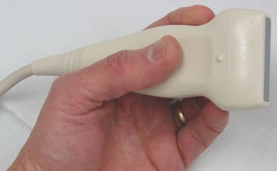
Using a process similar to sonar, a small probe generates an ultra-high frequency soundwave which reflects off the heart and is converted into an image of surprising detail. A series of images is put together to form a real-time video of your heart in action. When combined with technology designed to measure the doppler effect, (think of a changing sound a siren makes when it travels away from you), allows our machines to map the flow of blood inside the heart and let our physicians know if there are any places where the blood is flowing too abnormally, indicating a blockage or other abnormality.